)
Patients would potentially have earlier access to novel approaches.
German version/Deutsche Version
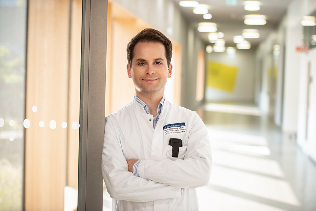
Medicine or computer science, that was the question Philipp Vollmuth asked himself when he was faced with the decision of which course of study to choose in 2006. He felt more drawn to medicine, but the hobby he had pursued most seriously during his school years was practical computer science. He had spent large parts of his free time programming. He decided to go into medicine but remained true to his enthusiasm for computer science during his studies in Innsbruck, especially since the possibilities of processing digital data with the help of learning machines were expanding rapidly during those years, and algorithms were making ever greater inroads into medical practice. The march of time therefore meant that Vollmuth’s interests and skills converged in an almost ideal way. Today, the Austrian heads the “Computational Neuroimaging” department he established at Heidelberg University Hospital. As a research physician, Vollmuth, née Kickingereder, works there at a particularly exposed interface of medicine and computer science, applying artificial intelligence (AI) techniques to the analysis of medical imaging. In the field of radiomics, he has quickly become one of Europe’s leading experts.
The first encounter with radiomics
During his doctoral studies at the Department of Neurosurgery at the University of Cologne, Vollmuth specialized in cancer research, or more precisely in the study of brain tumors. For three years, he researched “Stereotactic Interventions for the Diagnosis and Treatment of Brainstem Gliomas” there. The brainstem, which connects the brain to the spinal cord, controls vital functions such as breathing and heartbeat. It is also traversed by many motor nerves, so even the smallest tumors in it quickly cause paralysis. In most cases, they originate from glial cells, which are actually supposed to support and nourish the nerve cells. In stereotactic surgery, surgeons are guided in real time by tomographic images. How such images, which are also indispensable for the diagnosis and monitoring of brain tumors, could be captured and interpreted more precisely, began to occupy Philipp Vollmuth more and more. In 2012, a group of radiologists and oncologists from Maastricht presented a new approach to extract more information from medical images by informatic means: the radiomics approach.
The unearthed treasures of radiology
The name of this approach says it all: It demands that radiology should not be inferior to genetic research and molecular biology in the accuracy of its findings but should open up a complementary omics field by using the data it collects exhaustively enough. “It didn’t do that then,” says Philipp Vollmuth. “And it still doesn’t do so to a sufficient degree today.” Yet image data make up a large part of the data collected in healthcare and play a central role in treatment decisions. But still many radiologists rely primarily subjectively on their trained eye to assess an exam, and then measure the structures it depicts using conventional methods, rather than unveiling the wealth of data and turning it into information hidden in each image invisible to their daily routines. “In radiomics, we believe that medical images are not simply images, but contain a wealth of data.” This data can be used to derive conclusions about the best treatment option and make predictions about disease progression. However, this potential can only be tapped when images from many patients are linked together and evaluated according to strictly prescribed rules – in other words, according to an algorithm. First, the intensity of the images is normalized to make them comparable, then the tumor is identified and segmented, and finally radiomics features are extracted to quantitatively characterize its imaging phenotype. The most relevant of these data are incorporated into a predictive model, into which clinical and molecular data are also fed. The more individual data sets are available, the more the algorithm and model quality improve. “During this training, the system has to learn to distinguish what a tumor looks like on imaging,” Vollmuth says. “In subsequent tests with larger groups of patients, it then has to prove that it can actually distinguish.”
Artificial intelligence accelerates clinical trials
When Philipp Vollmuth took up a position in radiology and neuroradiology at Heidelberg University Hospital in 2013, he had no difficulties writing the programs and algorithms he needed to develop radiomic procedures himself. Although brain tumors continue to have an unfavorable prognosis, significant recent advances in their genetic and molecular characterization have yielded promising targets for targeted drugs. Some of these are already being tested in clinical trials. “Radiomics could relevantly accelerate such trials for the benefit of patients,” Vollmuth says. That’s because overall survival is poorly suited as a primary endpoint for clinical trials to treat rapidly progressing tumors, he says. It takes too long to obtain a statistically significant result. For the endpoint of progression-free survival, imaging is the decisive criterion. This is because it shows whether and when a tumor will grow again. Even in large multicenter studies, this decision must be made by a person responsible for it, who combines the findings of all radiologists involved. “However, in the manual assessment that has been used so far, radiologists do not always agree on the dynamics and progression timing of a tumor,” says Philipp Vollmuth. “When the tumor grows again and thus the endpoint of the study is reached is therefore often difficult and time-consuming to quantify.” In contrast, automated, objective reporting would be able to determine more precisely and quickly whether a new therapy is effective. “Patients would potentially have earlier access to novel approaches.”
Imaging instead of biomarkers
Vollmuth has already made numerous outstanding scientific contributions to AI-supported objectification of radiological diagnostics and prognostics of brain tumors. While still a resident, he became the first author of a prominently published study that retrospectively included 172 patients who had been diagnosed with recurrent glioblastoma by magnetic resonance imaging (MRI) and treated with bevacizumab at Heidelberg University Hospital between 2008 and 2015. This drug is a monoclonal antibody that inhibits the blood supply to tumors and may be used after unsuccessful standard therapy. However, there is as yet no validated biomarker that could predict whether a patient will respond to therapy with bevacizumab. Vollmuth showed that radiomics-extracted MRI data could in principle take on the role of such a biomarker. To do this, he mechanically identified 72 of each of a total of 4,842 radiomic features per patient that appeared to be particularly important in predicting treatment success – and validated that this was indeed the case. “Which drug we used to test this possibility is less relevant,” Vollmuth says. “Bevacizumab was just one example here.” [1]
Machine learning improves reporting
As head of the Computational Neuroimaging group, to which he was appointed in 2017, Vollmuth set out to demonstrate the power of AI for radiological diagnostics. To do so, he was able to draw on MRI data from 38 European hospitals in cooperation with the European Organisation for Research and Treatment of Cancer. He trained the AI algorithm with data from Heidelberg and then tested it with data sets from other hospitals. Among other things, he found that the AI-based three-dimensional quantification of tumor size had significantly better predictive power for tumor response to treatment than the two-dimensional Response Assessment in Neuro-Oncology (RANO) criteria previously used. [2] “To promote clinical translation of our approach, we made it available as applicable software in an open-source database.” For drug regulatory agencies, of course, this translation comes with a trade-off. For them to approve AI tools for use in routine clinical practice, they must be standardized as much as possible. On the other hand, AI tools are only useful if they keep learning and thus leave the originally agreed standard. “This is not currently resolved,” Vollmuth says. “However, the authorities are making efforts to map this phenomenon of continuously learning AI in regulatory terms.”
Strokes also targeted
Philipp Vollmuth is also exploring the possibilities of machine learning in stroke diagnostics as a cooperation partner in a European project. This project aims to enable faster diagnosis and, in particular, to support less specialized physicians and smaller hospitals in the diagnosis of strokes. By detecting vascular occlusions quickly and reliably through AI, physicians can make a correct diagnosis quickly and, if necessary, refer the patient to a specialized focus hospital for immediate therapy. The scientific project has the potential to improve stroke treatment and optimize patient care by providing efficient and accurate support for diagnosis and faster initiation of appropriate therapeutic measures. In the future, Philipp Vollmuth will be able to pursue his promising research work in the diverse spectrum of neuroradiology even more intensively, not only because of the Life Sciences Bridge Award of the Aventis Foundation. The Else Kröner-Fresenius Foundation has awarded him one of its Clinician Scientist Professorships in early 2023. Within the framework of this W3 endowed professorship to be established, Philipp Vollmuth intends to establish innovative AI-based solution approaches for radiological diagnostics and to enable their clinical translation.
Author: Joachim Pietzsch, Wissenswort
Photos: © Tobias Schwerdt
[1] Kickingereder P, Götz M, Muschelli J, Wick A, Neuberger U, Shinohara RT, Sill M, Nowosielski M, Schlemmer HP, Radbruch A, Wick W, Bendszus M, Maier-Hein KH, Bonekamp D. Large-scale Radiomic Profiling of Recurrent Glioblastoma Identifies an Imaging Predictor for Stratifying Anti-Angiogenic Treatment Response. Clin Cancer Res. 2016 Dec 1;22(23):5765-5771.
[2] Kickingereder P, Isensee F, Tursunova I, Petersen J, Neuberger U, Bonekamp D, Brugnara G, Schell M, Kessler T, Foltyn M, Harting I, Sahm F, Prager M, Nowosielski M, Wick A, Nolden M, Radbruch A, Debus J, Schlemmer HP, Heiland S, Platten M, von Deimling A, van den Bent MJ, Gorlia T, Wick W, Bendszus M, Maier-Hein KH. Automated quantitative tumor response assessment of MRI in neuro-oncology with artificial neural networks: a multicenter, retrospective study. Lancet Oncol. 2019