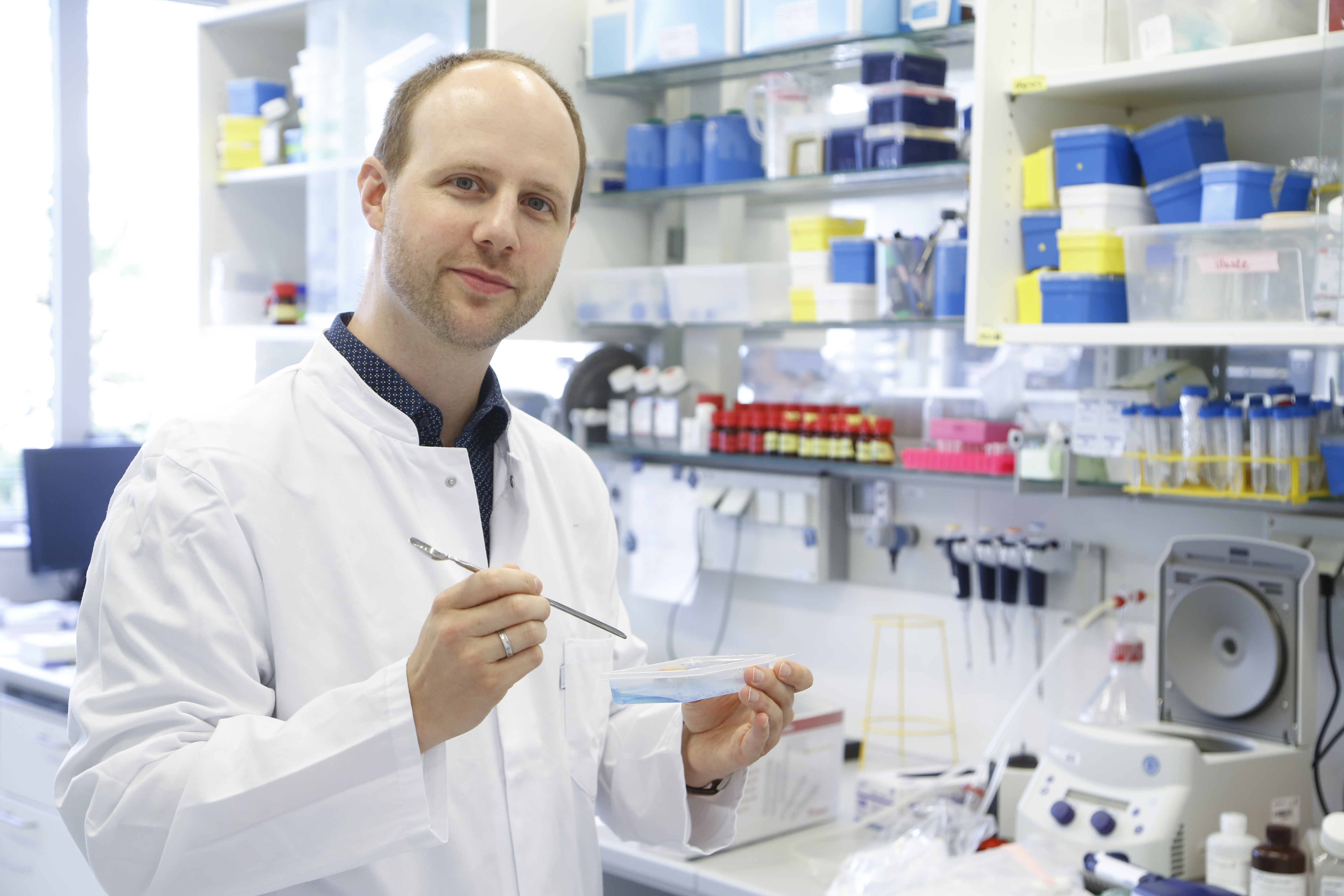
)
In general, we ask ourselves, how does a cell react when it experiences stress?
German version/Deutsche Version
If life were as simple as in a test tube, Christian Münch’s career would have been different. For in vitro, Christian Anfinsen had already demonstrated in the late 1950s that the three-dimensional shape into which a protein must fold in order to fulfill its function clearly results from the sequence of its amino acids – an achievement for which he was awarded a Nobel Prize in Chemistry in 1972. In a living cell, however, where molecules of all kinds crowd together in extreme confinement, this unambiguity can only be realized under the protection of molecular chaperones. These chaperones are there to prevent misfolding. They make up several percent of all intracellular proteins and are so numerous that all other proteins swim in them as if in a thick soup.
Chaperones for young proteins
The mass use of chaperones is vital because without their help, newborn proteins quickly go astray or are exposed to unprotected attacks. A protein is produced in a ribosome within a minute or so. When it emerges from its exit tunnel into the aqueous environment of the cytoplasm as an only slightly prefolded amino acid chain, it must find its correct spatial shape like lightning if it is intended for use in the cytoplasm. If it is intended for other sites of use, it is imported largely unfolded into the tunnel system of the endoplasmic reticulum (ER) or escorted to a mitochondrion for final folding. The environment of the cell is dangerous to an unfolded protein because half of all natural amino acids have hydrophobic side groups. If these are not quickly hidden inside the protein during the folding process, they clump together like fat droplets or with the hydrophobic groups of other proteins being formed. Many chaperones therefore already guard directly at the ribosome. They shield the hydrophobic part of a new protein until it can fold inward. They do this in a repeated cycle of grabbing and releasing until the whole protein is folded correctly. Other chaperones are shaped like a barrel in whose lumen a protein can fold undisturbed. This folding is not a chemical reaction, but takes place through the formation of numerous non-covalent bonds such as hydrogen bonds. Each protein should reach a state of as little free energy as possible along the way of this folding. Chaperones also help in this process. Because in the thermodynamic landscape that leads a young protein there, as if through a funnel of decreasing energy, there are chasms and dead ends from which it would not be able to escape on its own.
Characterized by the Munich triumvirate
Münch’s enthusiasm for protein folding in general and for the processes in the mitochondrion in particular was awakened in Munich. Although he studied biochemistry in Tübingen, his curriculum gave him the opportunity to spend a year in Munich, where he rotated through six different laboratories, including those of Walter Neupert, Franz-Ulrich Hartl and Stefan Jentsch. The encounter with this triumvirate became formative for his scientific career. Neupert was a world-renowned specialist in mitochondria, whose importance as metabolic centers would hardly be done justice if they were simply referred to as the power plants of the cell. Among his major discoveries was that mitochondria import proteins unfolded after they are made. In the late 1980s, he brought together in his lab Arthur Horwich, whom he had met at a conference, and his post-doc Franz-Ulrich Hartl – and both provided proof, defying the Anfinsen dogma, that the enzyme ornithine trancarbamylase can fold in the mitochondrion only in the presence of a molecular helper. They had thus discovered the first chaperone and founded the new research field of proteostasis, which deals with the dynamics of protein balance and its effects on health and disease. Stefan Jentsch investigated the quality control mechanisms used by cells in this process. He found out, for example, that and how unfolded proteins are exported from the ER and disposed of in the cytoplasm.
Almost like a prion
“Neurodegenerative diseases are mainly caused by protein misfolding,” says Christian Münch. “This was another reason why I chose a topic in this field for my doctoral thesis after completing my diploma in Tübingen.” He moved to the famous MRC Laboratory of Molecular Biology in Cambridge, England, to do his doctorate under Anne Bartolotti on a particular aspect of amyotrophic lateral sclerosis (ALS). He investigated the behavior of the enzyme SOD1, whose misfolding was known to be a central trigger of the pathogenic cascade leading to the onset of familial forms of ALS. Münch was able to demonstrate that the misfolded SOD1 spreads its mutated form rapidly like an avalanche, i.e. behaves like a prion, an infectious protein that induces changes in other proteins. It was “a revolutionary time” at the time, Münch recalls, because similar findings on various neurodegenerative diseases were published within a short time of each other. “My paper has been cited many hundreds of times.”[1] In 2011, Münch was awarded the British Neuroscience Association’s Postgraduate Prize for it.
A double answer
Equipped with a grant from the European Molecular Biology Organization, Münch joined Wade Harper’s group at Harvard Medical School in May 2012. The focus of his research there was on the special mitochondrial response to misfolded proteins, the mitochondrial unfolded protein response (UPRmt ). That living cells respond at all to the stress of a dangerously high amount of unfolded or misfolded proteins with a response to restore proteostasis had been discovered in the 1990s, first with respect to the ER, where all proteins destined for export and for anchoring in the cell membrane are folded, then for the cytoplasm and for the mitochondria, which have their own chaperones inside them like the ER. In principle, this response should always have two effects, namely to increase the production of chaperones and to temporarily reduce the production of other proteins. Abruptly, the supply of folding helpers can thus increase and the demand for folding decrease. From the ER and from the cytoplasm, this dual response is transmitted to the DNA in the cell nucleus after a reaction of special signal molecules with corresponding transcription factors. This could be worked out in detail by the early 2010s partly because good reagents existed for mammalian cell cultures that could be used to induce ER stress in the laboratory. For mitochondria, such highly specific reagents did not exist until then. Because it is surrounded by a double membrane, the mitochondrion cannot send its UPR into the nucleus as easily as the ER with the help of signaling proteins. Studying the UPR of the ER was particularly attractive to scientists because it became clear early on that diseases such as diabetes were clearly linked to ER stress. Mitochondria, on the other hand, have been treated rather stepmotherly, Münch says. “Only certain parts of them, such as the respiratory chain, have been heavily studied, other parts almost not at all.” Yet mitochondria are dysfunctional in the vast majority of diseases and are ultimately the organelles that trigger programmed cell death.
Extreme conditions in the mitochondrion
In any case, when Christian Münch started his work in Wade Harper’s lab, the UPRmt , unlike its ER and cytoplasmic counterparts, had only been fully described in the nematode C. elegans, but not in mammalian cells, where the situation is considerably more complex. Mitochondria have their own genome, consisting of 16,600 base letters, and encode mainly the 13 proteins essential for electron transport in the respiratory chain. However, a total of about 1,500 proteins are active in the mitochondrion, so a good 99 percent of them have to be imported. “These proteins are surrounded in the mitochondrial matrix by an extremely large number of highly reactive molecules that permanently damage them,” Münch says. Mitochondria could therefore probably actually fold and maintain proteins more robustly than other cell compartments. Nevertheless, the imbalance between correctly and incorrectly folded proteins, or not folded at all, that triggers the UPRmt often occurs in them. If one wants to study this particular UPR in cell cultures, one needs a substance that triggers it exclusively in mitochondria and does not simultaneously inhibit the very similar chaperones in the ER and elsewhere. And you have to do your subsequent measurements very quickly, because the direct effects of a UPR are soon overridden by compensatory effects.
Excellent solo effort at Harvard
Münch overcame both challenges in Boston. “He single-handedly developed new tools to explore the mitoUPR and made several discoveries in the process,” Harper praised his post-doc after the latter moved to the Institute of Biochemistry II of the Department of Medicine at Frankfurt’s Goethe University as group leader in 2016. That same year, a paper that only Münch and Harper had authored appeared in Nature. Such two-author articles are very rare: This is because they show that all the experiments described in them were undertaken by the first author alone. Münch had turned an inhibitor of the chaperone HSP90, which originated in cancer research, into a mitochondria-specific tool. “Mitochondria import proteins across a proton gradient in the membrane,” he says. “So I coupled the inhibitor with a strongly positively charged molecule so that it accumulates in mitochondria a thousand times more than in the rest of the cell.” Thanks to this trick, in sophisticated analyses of the transcriptome and proteome, he was able not only to intercept molecular messages that the mitochondrion sends to the nucleus in an emergency, but also to discover a previously unknown signaling pathway of the UPRmt through which the mitochondrion’s own genes are rapidly and reversibly inhibited in order to take “folding load” off it. [2]

Master of mass spectroscopy
Methodologically, Münch advanced to become an expert in quantitative proteomics with mass spectrometers. In Frankfurt, he expanded this expertise more and more. “We can look at eight to ten thousand different proteins simultaneously, basically all protein types of a cell.” To do this, the proteins must be taken from the killed cell, digested into peptide fragments and purified before they are shot into the spectrometer wrapped in an electrospray and read out in a differentially based on physical parameters. “With the latest equipment, we can even shoot in and measure 18 different samples at the same time.” In this way, the analysis of a cell culture produces a sequence of snapshots from which the time course of protein production during a UPRmt can be traced. In this way, “eavesdropping” on signal cascades can also succeed, since signals are predominantly transmitted via phosphorylations that can be detected by mass spectroscopy. This also applies to ubiquitination, which marks proteins for degradation.
Such degradation is the last resort when a protein can no longer be rescued by a UPR. “Then it’s done, that’s the life cycle,” Münch says, pointing out that in every healthy cell there is a constant interplay between misfolding stress and UPR. Each UPR is primarily protective. Only when the protein waste gets out of hand does it turn destructive. Then it provides not only for the disposal of proteins, but in the case of the UPRmt for the destruction of the affected mitochondria or even for programmed cell death. Understanding the “tipping points” between these stages is one of Christian Münch’s main concerns. “This is highly relevant for neurodegenerative diseases as well as for aging processes.”
The mitoUPR remains the center of attention
Because his laboratory is located in the university hospital, Münch’s basic research is closely linked to medical practice and can also draw on cell samples from patients. “We want to know in which diseases the mitoUPR occurs and by which biomarkers you can possibly detect it,” Münch says. “In general, we ask ourselves, how does a cell react when it experiences stress?” This can include stress caused by a lack of oxygen or aggressive oxygen radicals. “However, the mitoUPR is always our focus.” The importance of understanding it is evidenced not only by the Aventis Foundation’s Life Sciences Bridge Award but also by Münch’s multimillion-dollar grant from the European Research Council, which he has just applied to continue. The prospects for approval are good. That’s because recently Münch and his group resolved the question mark that had previously been in their account of the UPRmt . “We have discovered the unknown factor in the signaling chain from the mitochondrion to the nucleus.”
Author: Joachim Pietzsch, Wissenswort
Photos: © Uwe Dettmar
[1] Münch C, O’Brien J, & Bertolotti A. 2011. prion-like propagation of mutant superoxide dismutase-1 misfolding in neuronal cells. PNAS, 108(9):3548-53.
[2] Münch C & Harper J W. 2016. mitochondrial unfolded protein response controls matrix pre-RNA processing and translation. Nature, 534, 710-3.