)
That’s the beauty of basic research.
German version/Deutsche Version
When Roderick MacKinnon succeeded in elucidating the structure and functioning of a potassium channel in 1998, it was a scientific sensation. Just five years later, he was awarded a Nobel Prize. At this time, Inga Hänelt was studying biology in her seventh semester in Osnabrück. Her department was one of Germany’s strongholds of cell membrane research. Consequently, it was also a center for research into potassium channels. This is because the cell membrane, which consists of a double layer of fatty acids, is impermeable to potassium ions, as it is to other electrically charged molecules. Only through channels or with the help of active transport pumps can the vital ion pass through the cell membrane – and thus fulfill a condition for the transmission of electrical signals and the functioning of our nervous and cardiovascular system. The existence of such channels had already been proposed in 1925, and their neurophysiological significance was proven between 1950 and 1970. It was MacKinnon’s X-ray structural analysis, however, that provided insight into the precise molecular mechanism of a potassium channel for the first time.
Ion channels of bacteria are different
Although MacKinnon had made his findings on the potassium channel of a bacterium, they were transferred by researchers all over the world primarily to the situation in humans and other eukaryotic organisms, i.e. organisms equipped with cell nuclei, motivated by the hope that these findings could soon be put to medical use. Inga Hänelt, inspired by the microbiological excellence of her university, chose a different path. She focused her research on the transport of potassium ions on bacteria, chose the rod-shaped Vibrio alginolyticus, a relative of the cholera pathogen, as a model organism for her diploma thesis, and excelled in her dissertation, completed summa cum laude in 2010, with an investigation of its potassium channel KtrAB. This belongs to the “work animals,” explains Inga Hänelt, which under normal conditions ensure that bacteria take up enough potassium. “What drives me is that potassium transport in bacteria is very fascinating and at the molecular level quite different than in eukaryotic organisms.”
Within living cells, no positively charged ion is more abundant than potassium. Eukaryotic cells use a lot of energy to pump sodium out of and potassium into themselves. In this way, they establish an electrochemical gradient and create a membrane potential, i.e., an electrical voltage between the inside and outside of the cell. Electrical signals are transmitted by a temporary reversal of polarity and its rapid reversion. During this action potential, sodium channels first open and allow sodium to flow in. While these channels are still slowly closing, channels open through which potassium flows out and restores the original polarity. “Most biochemists think that potassium always flows out when a potassium channel opens, because that’s how they know it from neurobiology,” says Inga Hänelt. “In bacteria, however, it is usually different.” In them, the membrane potential is mainly generated by the flow of hydrogen ions (protons), which are pumped out through the electron transport chain in the course of cellular respiration. Under most conditions, the membrane potential thus established is sufficient for bacteria to allow potassium to flow passively into them against the concentration gradient by opening their potassium channels.
Potassium as a survival agent
Without this ability, bacteria could hardly survive. Without potassium, they would not be able to quickly adjust their metabolism to changing salinity, nutrient environments or acidity levels and thus adapt to almost all possible environments. Not only does potassium play a central role for bacteria in stabilizing pH and maintaining osmotic pressure and membrane potential, but they would also be unable to manage the regulation of their protein synthesis and the activity control of their enzymes without potassium. In contrast to multicellular organisms, which absorb environmental influences in a highly organized network of specialized cells, bacteria must immediately withstand the full force of their external environment. “How bacteria regulate their potassium balance with five or six different transporters and channels for this purpose is not yet entirely clear,” says Hänelt. “It’s not possible to deduce this from human structures.”
Potential targets for new antibiotics
Some bacterial potassium transporters do not occur at all in mammalian cells. There is much to be said for that they are pathogenicity factors, i.e. that a bacterium needs them to be contagious. “They give them intrinsic resistance,” Hänelt explains. “If bacteria didn’t have that resistance, they couldn’t survive in a human because they couldn’t handle the high salt concentrations they experience there.” Such bacteria-specific transporters, which have no homologs in humans, would in principle be suitable targets for new antibiotics. “This is highly topical. Studies estimate that by 2050, more people will die from bacterial infections than from cancer.”
The potassium channel KtrAB could become such an antibiotic target. It ensures the well-being of a bacterium under moderate environmental stress. The letters “A” and “B” denote its two main parts comprising 230 and 455 amino acids, respectively. “B” consists of two nearly parallel channel shafts whose walls bore helically through the membrane. “A” consists of eight subunits grouped into an oval ring that abuts the membrane from inside the cell. When the potassium level is sufficient, this ring is linked to ADP molecules. In this ring are two connectors resembling the blades of two jackknives, the shaft of each of which lies in one of the channel wells. The mechanical tension of this connection presses the channel walls together and closes them to the passage of potassium ions. However, if their intracellular concentration falls below a certain level, the bacterium produces the more energy-rich ATP as if in panic. This displaces ADP from the “A” ring and abruptly deforms it into a square, causing the knife-blade-like connection to snap back to the channel shafts and a flood of about a billion potassium ions per second to pour through the two shafts.
A pump for emergencies
When Inga Hänelt discovered this mechanism and published it for the first time in 2016[1], she had already been conducting research at Goethe University in Frankfurt for several years. After completing her doctorate, she had initially left Osnabrück for Groningen, equipped with a two-year fellowship from the German Research Foundation. There, in 2012, she received an offer from Frankfurt, the “Europe-wide center for the structure-based biochemistry of membrane proteins,” as she puts it, to join the Department of Molecular Microbiology and Bioenergetics. She quickly succeeded with her work on transport and communication processes through biological membranes and in 2015 simultaneously took over an Emmy Noether junior research group in the Institute of Biochemistry with a junior professorship, in which she turned her attention to the potassium pump KdpFABC in addition to the potassium channel KtrAB. This pump only starts up in an emergency of extreme potassium shortage – and may only be formed and active in this case, because its force is so great that otherwise potassium poisoning would be the result. Therefore, only when the potassium concentration is extremely low does a potassium sensor anchored in the membrane send the command to produce the pump to the bacterial genes responsible for it. In the process, the pump proteins are provided with phosphate groups after their production. This integrates them into an intracellular signaling network that ensures that the existing KdpFABC pumps are switched off as soon as the potassium level is balanced again.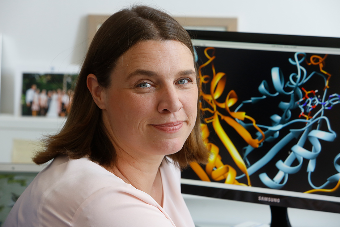
Highly complicated small machines
“Don’t touch that, this protein behaves so badly!” colleagues warned her when she started working on the pump, Hänelt recalls. But she wasn’t put off by that, she says, because she was fascinated by the chimeric character of the pump, one subunit of which resembles a channel. In fact, KdpFABC “behaved incredibly well,” so that she and her group were able to quickly elucidate its architecture.[2] For a structural biologist, good behavior means that a protein can be detached from the membrane undamaged and prepared in a realistic manner for examination purposes. After all, a potassium transporter is not just a blob of protein, but a highly complicated little machine with many moving parts and joints that interlock in well-ordered sequence to do their job. “They’re happy in their membrane and as soon as I take it away from them, the problem starts to keep them stable.” When X-ray crystallography was the predominant method for determining structure, the main difficulty was getting a protein into crystal form. Today, when cryo-electron microscopy (cryo-EM) dominates, the key is to cool a protein to below minus 150 degrees Celsius in a fraction of a second, and isolating the functional protein is the biggest challenge.
Interbacterial communication via electrical signals
The signal molecule cyclic di-AMP (c-di-AMP) has increasingly become the focus of their research. In Gram-positive bacteria, it serves the overriding regulation of potassium balance by simultaneously modulating all transport pumps and channels as needed to increase ion export and decrease ion import. Gram-negative bacteria do not appear to produce c-di-AMP, but probably employ a similar super-regulator. To date, it has not been identified. Not all potassium channels, however, respond to c-di-AMP. Among them is probably the channel YugO, which is significantly involved in an astonishing phenomenon discovered in the gram-positive Bacillus subtilis, namely in the communication of different bacteria by means of electrical signals. It has been known for decades that bacteria communicate with each other because they can only achieve their goals together. Until now, the main language in which they do this was considered to be quorum sensing. In relatively complicated chemical words, they exchange information, for example, about when they have reached a sufficient number to be able to attack as safely as possible or to form a resistant biofilm. Hänelt is convinced that all bacteria also talk to each other via electrical signals, through the exchange of simple rapid sequences of letters in the form of ionic currents, comparable to the transmission of information via nerve conductors in complex multicellular organisms. “There are already therapies today that involve applying dressings to wounds that generate a current, thereby stopping the growth of bacterial biofilms. We just don’t yet understand exactly why that works.”
Basic research needs good cooperation
Solving this puzzle requires bridging the gap between structural biology and microbiology. There is no other way to understand experimentally how many bacteria interact in a biofilm. Inga Hänelt succeeds in building this bridge thanks to a dense network of research collaborations that she has established, true to her motto “It is better to work together than against each other.” One such collaboration is the collaborative research center “Protein networks and machinery in cell membranes” established at Goethe University in 2022. In it, for example, she uses the optogenetic expertise of friendly laboratories to gain electrophysiological insights. Further promising perspectives could be opened up by a cluster of excellence that Hänelt is currently applying for together with other Frankfurt researchers. In it, subcellular structures are to be considered in their natural context. “We are trying to cut entire boxes out of a cell and find out what is happening inside.” Cryo-electron tomography, which is still in its infancy, should be particularly helpful. It may be possible to look at the molecular events in an entire bacterium in this way.
Since 2021, Hänelt has held a Heisenberg professorship at the Institute of Biochemistry at Goethe University. Support from the Aventis Foundation’s Life Sciences Bridge Award has contributed to this. The permanent position gives her the security of being able to continue her research without fear for the future. She also hopes to gain more precise insights into the potassium channel KtrAB, for example. Because its opening mechanism is more complicated than thought. The potential target for future antibiotics is “the most difficult protein to understand that we are working on,” says Hänelt. And she says it with joy. “In industry, it would have long been said: leave KtrAB, we’ll make money faster with another project. At the university, that’s not a criterion. That’s the beauty of basic research.”
Author: Joachim Pietzsch, Wissenswort
Fotos: © Uwe Dettmar
[1] Diskowski, M., Mehdipour, A.R., Wunnicke, D., Mills, D., Mikusevic, V., Bärland, N., Hoffmann, J., Morgner, N., Steinhoff, H.J., Hummer, G., Vonck, J.#, Hänelt, I.# (2017) Helical jackknives control the gates of the double-pore K+ uptake system KtrAB. eLife, 6:e24303.
[2] Stock, C., Hielkema, L., Tascon. I., Wunnicke, D., Oostergetel, G., Azkargorta, M., Paulino, C.#, Hänelt, I.# (2018) Cryo-EM structures of KdpFABC suggest a K+ transport mechanism via two inter-subunit half-channels. Nat. Comm., 9:4971