)
Observing the basic components of life with my own eyes.
German version/Deutsche Version
X-ray crystallography was the undisputed queen of structural biology in the 20th century. It is thanks to her that we now know how genetic information is stored, transcribed and translated into the language of proteins; under her aegis, the structures of the molecules and molecular machines that determine the flow of life, the structures of DNA, RNA polymerase and the ribosome, were decoded. But no one remains queen forever. The deeper science delves into the dynamics of life, the more clearly crystallography is reaching its limits. New methods have long since taken over as sidemasters, nuclear magnetic resonance spectroscopy (NMR) of course, but also cryo-electron microscopy (cryo-EM) and cryo-electron tomography (cryo-ET). They are Stefan Pfeffer’s specialty, in which he has developed a mastery over the years that step by step opens up previously unknown insights, especially into processes related to the work of the ribosome, that giant production factory for proteins that is one of the largest cellular structures and occurs a million times in some cells of our body.
Beyond the boundaries of crystallography
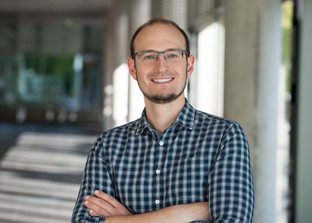
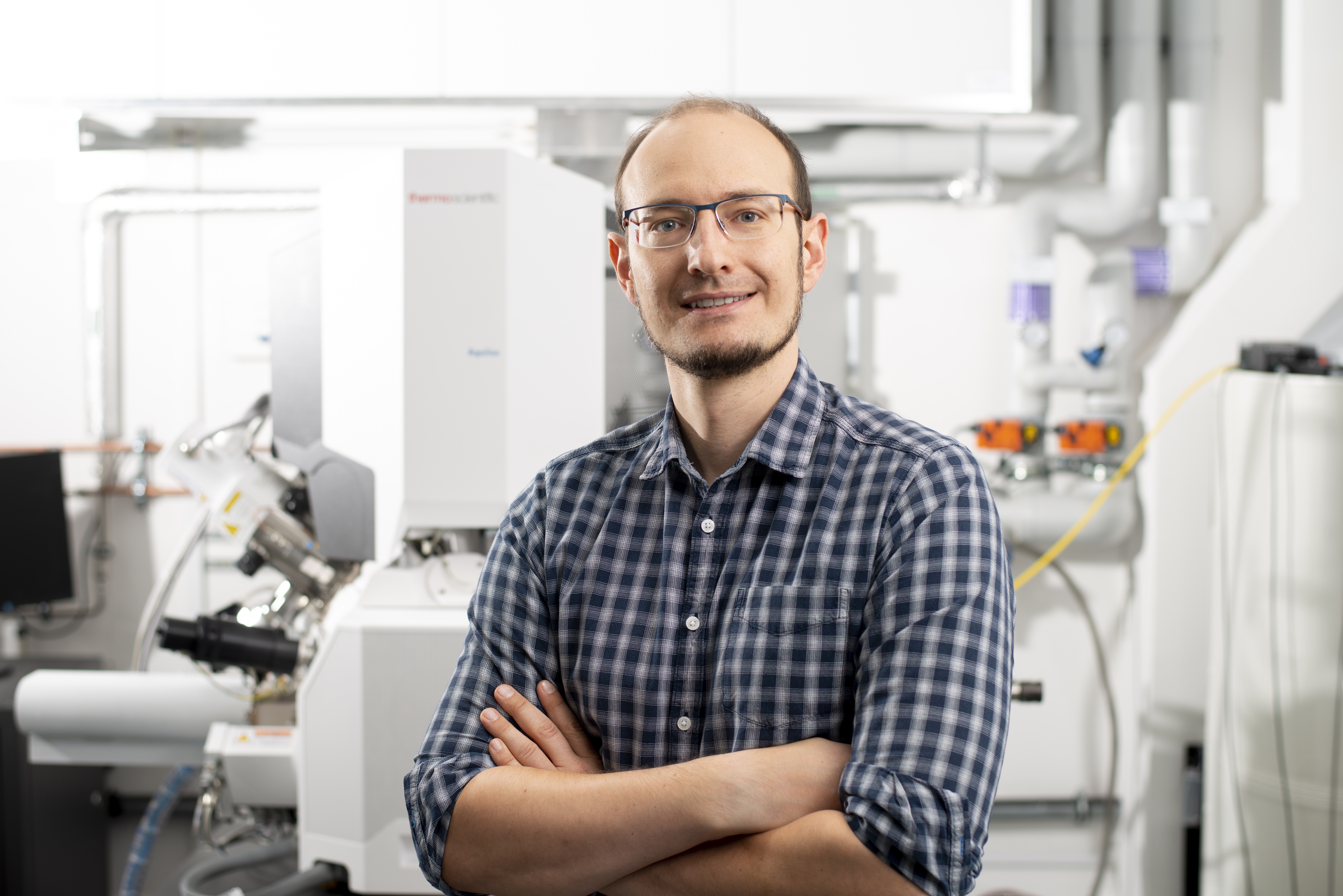
“Structural biology has always fascinated me,” says Stefan Pfeffer, who already researched ribosome-associated processes in his diploma thesis and now heads a junior research group at the Center for Molecular Biology at Heidelberg University (ZMBH). “Observing the basic components of life with my own eyes, so to speak, is very appealing to me because it’s in stark contrast to what a biochemist normally sees in the lab.” In addition, he said, it is the structure, surface and mobility of larger macromolecules that drive biochemical reactions. This relationship between form, motion and function can no longer be adequately explained by the tools of X-ray crystallography alone. This is because it is based on the fact that X-rays are diffracted in crystals like waves, so that the structure of the respective crystal can be calculated from the interference pattern of the diffracted rays. So proteins must be crystallized for their structure to be revealed by X-rays. “That’s the hurdle that’s difficult to overcome because many proteins are too flexible to be crystallized.” But if crystallization succeeds, the protein’s atoms are constrained in a lattice that deprives them of freedom of movement and usually allows only a single still image. Cryo-EM does not have these two limitations. In principle, all proteins in moving action are accessible to it. “Because we keep our samples in aqueous solution in cryo-EM, the proteins map the full range of their physiological motility after they have been fixed by rapid freezing. We have computational approaches to filter out the different conformations and analyze them separately,” Pfeffer explains. Ideally, these are movements that proceed in discrete steps, i.e., where components of the complex are located at precisely defined points at specific times. This possibility of extracting snapshots of a movement from a data set and then stitching them together partially compensates for the disadvantage to NMR, for which dynamic processes in aqueous solution are directly accessible, but with reasonable effort only for relatively small proteins.
Picture puzzle with ribosomes
The view of the entire ribosome thus eludes NMR, but offers a prime example of the visual power of the two cryo techniques. “The ribosome goes through a great many conformational states as it does its work,” says Stefan Pfeffer. “And with cryo-tomography in particular, we can track these states in a trajectory of motion within intact cells.” The ribosome is a complex of ribosomal RNA and proteins that performs two main tasks to make cellular proteins. It decodes the genetic message of messenger RNA (m-RNA) coming from the nucleus, and it makes peptide bonds between amino acids, which correspond to that message and are delivered by transfer RNA. The ribosome consists of a large and a small subunit. In resting phases, they are separated from each other. They assemble at the start codon of an mRNA only when translational work begins. As they thread the mRNA between them, they link about four amino acids per second, stopping when a stop codon appears. The resulting protein chain leaves the ribosome through an exit tunnel in its large subunit. The message of an mRNA is usually not exclusive to one ribosome: Dozens of ribosomes often attach to the same mRNA within a short distance of each other. “These polysomal arrangements play a crucial role in the successful production of proteins by keeping the nascent peptide chains separate from each other, allowing them to fold undisturbed.” That’s necessary, too, because, like in a picture puzzle, ribosomes are so densely packed in the cytoplasm that they account for at least half of its volume.
Explorations in the New Territory of Cryo-Electron Tomography
For all cryo-EM methods, the objects to be examined must be frozen in their natural state. The cooling rate must be so high that the water present in and around the sample does not form ice crystals, but remains in a completely disordered state. “We achieve this with vitrification robots that freeze the sample very quickly in liquid ethane, which has a very high thermal conductivity,” Pfeffer explains. During the subsequent experiments, a temperature of minus 150 to 160 degrees Celsius must not be exceeded to prevent ice crystals from forming, which would destroy the sample. “This requires a delicate touch when handling the frozen samples in liquid nitrogen.” Largely due to the development of cameras that detect electrons directly without having to convert them temporarily into a light signal , classical cryo-EM is now almost a routine procedure in research. It can determine the structure of individual proteins at nearly as high a resolution as X-ray crystallography. In 2017, its development was crowned with the Nobel Prize in Chemistry. In contrast, cryo-ET is an area whose exploration is far from complete. “It provides us with a 3-D image of the entire macromolecular landscape of a cell,” says Stefan Pfeffer, who has been involved in the exploration of this landscape since completing his doctorate at the Max Planck Institute of Biochemistry in Martinsried.[1]
The fight against background noise
An MRI scanner can capture a patient’s organs layer by layer and construct images from them. A cryo-electron microscope cannot do that with a shock frozen cell, because an electron beam can only shine through samples less than 0.3 millionths of a meter thick. “However, manual cutting is out of the question with a frozen cell, which must not thaw and should not deform during cutting.” Instead, the specimens are created in the Cryo-ET by cryo-focused ion beam milling. In this process, the samples are scanned with a focused beam of high-energy gallium ions. Layer by layer, the molecules of the cell thereby pass into the gas phase and are removed with a vacuum pump without heating the rest of the cell. “This is how we ablate 95 percent of the cell,” Pfeffer says. “For tomographic examination, only a single thin frozen lamella of each cell remains, which is then imaged three-dimensionally at molecular resolution using Cryo-ET.” The main challenge is to tease out specific structures from these data, like mineral resources from a mine. Much more significant than for classical cryo-EM, which relies on mathematical averaging of many hundreds of thousands of particle data, is the data analysis for cryo-ET. “As with cryo-EM, we have to transform the two-dimensional images in our raw data into spatial views, and we have to factor out a lot of noise in the process,” says Stefan Pfeffer. “This is very difficult in cryo-ET because the signal is quickly lost in the background noise due to the thickness of the sample.” He therefore also devotes a large part of his creativity to optimizing these processes. Yet the resolution of cryo-ET is still limited. For this reason, Pfeffer relies on the complementarity of both approaches. He uses cryo-EM to produce structures of individual proteins at near-atomic resolution, and then uses cryo-ET to study their interaction with other cellular components.
At the birthplace of proteins
This complementary approach has particularly high prospects of success with ribosomes because they are the most abundant and nearly the largest macromolecular complexes in a cell. For example, it offers the chance to follow exactly how the birth of a protein occurs as it leaves the exit tunnel of a ribosome, which is initially so narrow that the backbone of the just-linked amino acid chain with its side groups just fits through, but then widens into a vestibule that gives the chain a free space for initial helical and sheet-like folding. “As long as the nascent protein is still in the ribosome, we can see it very well; after that, it escapes our view.” As soon as it emerges, it strives to gain its correct shape as quickly as possible. Otherwise, it would run the risk of clumping with other new-born proteins. To prevent this, the tunnel exits on the polysomes are spatially separated to a maximum, says Pfeffer. Together with his research group, he has succeeded in elucidating the structure of a homolog of one of the most important enzymes at the exit of that tunnel. This enzyme is methionine aminopeptidase (MetAP).[2] By default, the synthesis of any new protein begins with the amino acid methionine; accordingly, it leaves its birth canal first. MetAP is responsible for splitting it off again from many proteins. Thus, this enzyme is one of the signals that are integrated at the tunnel exit to decide on the further molecular fate of a nascent protein, explains Stefan Pfeffer.
How cellular stress shakes up structures
Receiving the Aventis Foundation’s Life Sciences Bridge Award has raised awareness of his outstanding achievements. He was recently awarded an ERC Starting Grant – for a project in which he is studying how ribosomes respond to processes that hinder the folding of new proteins or cause already folded proteins to clump together. “Such processes are directly linked to neurodegenerative and also neuromuscular diseases.” Experimentally, Pfeffer establishes these stress situations, for example, through heat-mediated clumping of proteins within cells, or through manipulations of the translational translation process that specifically lead to the production of unfinished proteins. Cells respond to such stress primarily by two actions: They shut down general protein synthesis to prevent the misfolding of newly formed proteins as much as possible, but at the same time they increase the synthesis of chaperones. These are those proteins whose specific job is to help young proteins fold properly. “On a biochemical and cell biological level, these processes are relatively well understood, but how they affect the structure and molecular organization of ribosomes within the cell is still largely unmapped territory,” says Stefan Pfeffer. From cryo-electron tomographic exploration of this terrain, he hopes to gain insights into physiological and pathological processes that molecular biology has not yet found – and to discover new approaches that will lead to the development of better treatment options for diseases triggered by protein misfolding.
Author: Joachim Pietzsch, Wissenswort
Photos © Tobias Schwerdt
[1] Cf. Pfeffer, S.*, Woellhaf, M.W.*, Herrmann, J.M., Förster, F., 2015. Organization of the mitochondrial translation machinery studied in situ by cryoelectron tomography. Nat Commun 6, 6019.
[2] Wild, K., Aleksic, M., Lapouge, K., Juaire, K., Flemming, D., Pfeffer, S°, Sinning, I.°, 2020. MetAP-like Ebp1 occupies the human ribosomal tunnel exit and recruits flexible rRNA expansion segments. Nat Commun 11, 776.