

)
If this organization can be altered in a targeted way, that is a new point of attack for drugsGerman version/Deutsche Version
A Scholarship Foundation Blazes the Trail
After finishing high school in Bamberg, Leonhard Möckl knew that he wanted to study chemistry, but not whether he was drawn more to classical chemistry, to its biological applications, or to its physical aspects. The “Y model” at LMU Munich suited him well, since it allowed him to study chemistry and biochemistry in an integrated fashion before specializing. “That equipped me from the very beginning with an interdisciplinary mindset, which greatly benefits my research today.” It was thanks to the Studienstiftung des deutschen Volkes (the German Academic Scholarship Foundation), which accepted him in 2009 at the end of his first semester, that Möckl nonetheless found an early focus. The foundation offers its scholarship holders the chance to take part in academic colloquia that spend two years working intensively on an interdisciplinary topic within their field. When he saw the announcement for a colloquium titled “The Sugar Code,” led by Thisbe Lindhorst, a professor in Kiel, he was somewhat bewildered, Möckl recalls, because he had never heard of glycobiology. “After the first sessions of the colloquium I was, on the one hand, even more bewildered, because I realized how much I did not know. On the other hand, I was fascinated and thought: this is knowledge I want to acquire, and this is what I would like to do research on.”
The Glycocalyx Eludes the Eye
That was the spark for Möckl’s rapid ascent into the avant-garde of glycocalyx researchers. Pioneers of cell biology such as George Palade were once unable to say with certainty whether this structure was merely an artifact. Under the electron microscope they had seen a fluffy coating on the surface of the endothelial cells that line our blood vessels, and at first suspected that it might be nothing more than a precipitate produced during specimen preparation. In reality, it was a structure that had been made to collapse during preparation. The glycocalyx consists largely of water. In its natural state it protrudes into the vessel lumen in a swollen condition. As soon as a specimen is prepared for electron microscopy by conventional methods and dehydrated, it collapses in on itself. With the dye ruthenium red, which attaches to negatively charged sugar residues, it was at least possible in the mid-1960s to visualize it for the first time in its collapsed state. It took another 30 years, however, before intravital microscopy made it possible to estimate the thickness of the glycocalyx in the capillaries of hamster muscles with reasonable accuracy at half a micrometer. Today we know that it can be far thicker in large vessels. Even though endothelial cells are the traditional object of glycocalyx research because they are easily accessible, all cells—including those located relatively far from blood vessels—carry a sugar coat as well. Deciphering its structure with ordinary light microscopy, however, is possible only to a limited degree. Its resolution limit is around 250 nanometers.
The Sweet Side of the Membrane
Möckl’s most important mentor in Munich was Christoph Bräuchle, who held the chair of physical chemistry at LMU. Under his supervision Möckl wrote a bachelor’s thesis that impressed Bräuchle so much that he offered him the opportunity to pursue his doctorate and his master’s thesis in parallel, immediately afterwards and under his guidance. “Actually, I had wanted to leave Munich for my master’s degree, but I could not turn down that offer,” Möckl recalls. Bräuchle gave him a great deal of freedom and extended considerable trust in advance on a project that was initially very unclear. He told him: “If these sugars interest you that much—I myself know little about them, but I will support you if you bring in additional expertise.” And so it came about that Bräuchle became his doctoral father and Lindhorst his doctoral mother, jointly supervising his thesis “The Sweet Side of the Membrane—Sugars in Biophysics, Bacterial Adhesion, and siRNA Delivery,” which he completed summa cum laude in 2015. In it, Möckl says, he investigated many dynamic processes through glycobiological questions, but he ran up against the limits of the established methods. That motivated him to learn the technique of high-resolution single-molecule light microscopy as a postdoctoral researcher.
Two Award-Winning Mentors
Möckl summoned his courage and applied to the laboratory of W. E. Moerner at Stanford, who as one of the inventors of this technique had been awarded the Nobel Prize in Chemistry in 2014. In the aftermath of that honor, however, Möckl’s application initially got lost in Moerner’s inbox. But Christoph Bräuchle encouraged him to follow up after some time had passed. That led, in 2016, to a two-and-a-half-hour Zoom conversation between Moerner and Möckl, and shortly thereafter Möckl moved from Munich to Stanford. By then, Carolyn Bertozzi had also moved there from Berkeley—an outstanding glycochemist who had transferred the principle of click chemistry to living cells and who would go on to share the 2022 Nobel Prize in Chemistry with the inventors of that principle. In this environment, Möckl’s interdisciplinarity proved to be a hinge: “I understood less about glycobiology and click chemistry than Carolyn, and less about single-molecule microscopy than W.E., but at some point, I understood enough of both to bring them together.” Indeed, under the supervision of Moerner and Bertozzi, Möckl succeeded—by combining the two methods—in showing for the first time on the nanometer scale how the glycocalyx is organized. On that occasion he additionally showed that cancer cells expand their glycocalyx, probably in order to camouflage themselves from the immune system.1
Super-Resolution Plus Click Chemistry
When two molecules lie closer together under a classical light microscope than half the wavelength of light, their signals overlap and merge into a blurry spot. The super-resolution microscopy that Moerner helped develop circumvents this limitation by ensuring that molecules positioned too close to one another do not emit light at the same time. They are equipped with fluorescent markers that can be switched on and off. In thousands of images recorded one after another, different molecules of the specimen light up depending on the state of the switch. Their individual positions can then be calculated. Finally, by superimposing all the individual exposures, one obtains a “super-resolved” overall image. The problem Möckl faced, however, was this: unlike proteins, sugars cannot be furnished with genetically encoded fluorescent switches such as GFP, because “sugar coating” is the result of post-translational modifications. Falling back on labeling with immunofluorescence was ruled out as well, because antibodies bind to sugars too unspecifically on the one hand and can produce mislabeling that distorts the result on the other. He solved this problem with the help of click chemistry. Its best-known example: two organic substances—one containing at least one triple bond between two carbon atoms (an alkyne), the other three linked nitrogen atoms (an azide group)—join together almost as quickly as the two halves of a snap fastener, with a click; copper, however, serves as the catalyst. Bertozzi had modified this procedure and thereby made it usable in a detoxified form for applications in biological research. Möckl benefited from this in his glycocalyx project: he fed living cells sugars to which he had previously attached azide handles. The cells incorporated these sugars into their glycocalyx. Their azide handles could now easily be coupled to fluorescent dyes by means of click chemistry. Möckl determined the height of the glycocalyx—whose structure he was now able to analyze by examining various cell cultures under the microscope—using software he had written specifically for this purpose.
The Spirit of Stanford
“The experience of working patiently on something and, in doing so, letting something truly good emerge—I lived through that more intensely at Stanford than ever before,” says Leonhard Möckl. “Much of what I accomplished at Stanford I would not have accomplished elsewhere; the atmosphere carried me.” The highly competitive environment gave him wings. “It is stimulating in a way that is hard to put into words when almost every person around you is excellent in his or her own way.” There was, he says, a “constant openness to new ideas.” The basic attitude at this elite university, he explains, was: “You can do whatever you want, and we will support you.” That is how he was able to teach himself machine learning at a time when the field was still very young, and it is also why he was admitted as a matter of course to the advanced philosophy seminar run by Robert Harrison and Sepp Gumbrecht, in which he took part for three years until the pandemic brought it to a halt. When Möckl moved to the Max Planck Institute for the Science of Light in Erlangen as a research group leader in 2020, he brought with him, along with Californian verve, the confidence that he would soon be able to zoom deeper into the glycocalyx. “As fine as the 2019 publication is, we had to average over many signals in it and did not push through to the level of individual sugars. The resolution was not yet sufficient for that.”
A Researcher with a Gift for Combination
That his confidence was justified was something Möckl demonstrated with his Erlangen team in 2025.2 Less than one millionth of a millimeter—those are the distances between the sugars of the glycocalyx that he can now measure and visualize. This, once again, is the result of his brilliant knack for tailoring and combining the very latest methods precisely to the question he wants to answer. To advance into the ångström range of the sugar coat, he applied two techniques that Ralf Jungmann had developed in Munich: DNA-PAINT and RESI. “That turned out to be the key to reaching these high resolutions.” Unlike the super-resolution microscopy pioneered by Moerner, Jungmann’s approach does not use switchable fluorescent dyes but DNA probes, which make the observed molecules blink in different colors. With RESI, these signals are then read out in a large number of sequential rounds and assembled into a pattern. That was an enormous task for his entire team, Möckl says. “On behalf of everyone, I would like to thank my first doctoral student, Karim Almahayni. He carried out the optimization experiments with incredible motivation—with methods like these, everything simply has to be right. Because there are so many parameters that you are essentially blind if even one of them is off. Karim’s contribution was decisive.”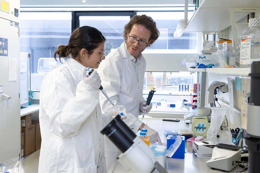
A Teacher with Depth
Supporting the next generation of scientists is a concern for Leonhard Möckl outside his main job as well. Since 2023 he has led workshops each year at the Lindau Nobel Laureate Meetings, together with council member Pernilla Wittung-Stafshede, in which the Lindau Guidelines for a sustainable scientific culture are further developed with the Young Scientists from all over the world who attend. “The guidelines are formulated in very general terms,” Möckl explains. “That is intentional, but the question is how they can be implemented in practice. One guiding principle, for example, reads: ‘Foster all talent.’ But what does that mean in an appointment procedure, say? After all, I cannot decide in favor of every candidate. Reflecting on and describing this leap from the idea to its realization is the aim of our workshops.” Regularly reflecting on what he does scientifically is also part of Möckl’s life as a professor. “In my most successful course, I deal with the logical foundations of scientific knowledge. In it we work through the great milestones in the philosophy of science.”
On the Way to a Milestone?
One of the milestones of molecular biology could one day be an atlas in which the spatial arrangement of sugar structures on the cell surface is systematically mapped and differentiated by cell type. Möckl has set out to reach this milestone. He reports on the distance he and his team have covered so far in a paper that Nature Nanotechnology has accepted for publication.3 In it he has worked out the topography of the glycocalyx of healthy cells as well as of breast cancer cells, nerve cells and immune cells. He has correlated it with the condition of these cells and recorded how it changes and under which conditions. In doing so, he found confirmation, for example, that cancer cells hide in an ever-thicker sugar coat the further they progress. But he also determined that T cells restructure their glycocalyx within minutes once they have been put on alert. “The clinical implications are obvious,” says Möckl. “If the spatial organization of the glycocalyx reflects the state of a cell, that is a new variable for diagnostics. If this organization can be altered in a targeted way, that is a new point of attack for drugs.” Everything therefore suggests that it is worth continuing along the path he has embarked upon. The Aventis Foundation intends to support him in this endeavor with its Life Sciences Bridge Award.
Author: Joachim Pietzsch, wissenswort
Photos: © Uwe Dettmar
Notes 1 L. Möckl*, K. Pedram*, A. R. Roy, V. Krishnan, A.-K. Gustavsson, O. Dorigo, C. Bertozzi, and W. E. Moerner, “Quantitative Super-Resolution Microscopy of the Mammalian Glycocalyx”, Dev. Cell 50, 57 (2019), https://doi.org/10.1016/j.devcel.2019.04.035 2 K. Almahayni*, L. Masullo*, I. Pachmayr, M. Honsa, L. Heinze, S. Fritsche, H. Grabmayr, R. Jungmann, and L. Möckl, “Ångström-resolution imaging of cell-surface glycans”, Nat. Nanotechnol. 20, 1457 (2025), https://doi.org/10.1038/s41565-025-01966-5 3 D. M. Joseph,* N. Yurekli,* S. Fritsche, R. Hashem, O.-M. Thoma, I. Larafa, T. Boric, C. Bielawski, K. Almahayni, K. Franze, M. Waldner, and L. Möckl, “Glycan Atlassing: Nanoscale analysis of glycocalyx architecture enables functional tracing of cell state”, accepted for publication at Nat. Nanotechnology. Preprint: https://www.biorxiv.org/content/10.1101/2025.04.29.651200v1